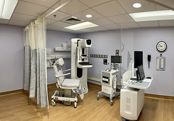
Picture of room with 3D mammography equipment.
Imaging Services/Nuclear Services
Ashe Memorial Hospital is proud to offer some of the most advanced imaging services available. These tools help medical providers quickly assess your medical issues and begin treatment promptly. To learn more about any of the tests below, or to schedule an appointment, call 336-846-0820.The imaging services offered at Ashe Memorial Hospital include:
Nuclear Medicine
Nuclear medicine is an imaging modality that uses small amounts of radioactive material combined with a carrier molecule. This compound is called a radiotracer or radiopharmaceutical. Doctors use nuclear medicine tests to diagnose and/or treat various diseases. These include cancer, heart disease, gastrointestinal, endocrine, and neurological disorders. They are non-invasive and usually painless.
CT Scans
CT is short for Computed Tomography. Commonly called CT or CAT scan, Computed Tomography is the preferred modality for many conditions and situations. The Emergency Room (ER) commonly uses CT because the exams are done quickly and can be used to evaluate multiple organ systems at one time. CT works by using X-rays and controlled motion to make a digital representation of the body. That digital representation can then be displayed as images from top to bottom, side to side, or any way in between. CT can be used to show any part of the body – particularly lungs, bones, organs, and blood vessels.
Ashe Memorial Hospital uses a state-of-the-art GE 128 Revolution Maxima CT scanner. This technology offers shorter scan times while using a personalized, low patient radiation dose approach. CT can generate images of internal organs, bones, soft tissue and blood vessels, producing views inside the body similar to a loaf of bread.
Ultrasound
Ultrasound (also called sonography or ultrasonography) is a noninvasive imaging test. An ultrasound picture is called a sonogram. Ultrasound uses high-frequency sound waves to create real-time pictures or video of internal organs or other soft tissues, such as blood vessels.
Ultrasound enables ultrasound technologists to “see” details of soft tissues inside your body without making any incisions (cuts). And unlike X-rays, ultrasound doesn’t use radiation.
Although most people associate ultrasound with pregnancy, healthcare providers use ultrasound for many different situations and to look at several different parts of the inside of your body.
Mammography
One of the most powerful tools in the diagnosis and treatment of breast cancer, mammography creates a digital image of tissues within the breast.
Stereotactic Breast Biopsy
Stereotactic breast biopsy is a form of image-guided biopsy that uses mammography to help your radiologist locate an abnormality, then guide a core-sampling needle to the area to remove a small amount of tissue. This tissue sample is then sent to a lab for analysis.
Magnetic Resonance Imaging (MRI)
An MRI (Magnetic Resonance Imaging) is a non-invasive medical imaging technique that uses a large magnet, radio waves, and a computer to produce detailed images of the inside of the body. Unlike CT scans or X-rays, MRI does not use ionizing radiation. Before the scan, patients may need to remove metal objects and possibly fast if imaging your abdomen. It’s crucial to inform your scheduler if you have any metal in your body, such as pacemakers, implants, or metal fragments including old gun wounds, as these can affect your eligibility for the scan.
During the MRI, you’ll lie on a table that slides into a tunnel-shaped machine and you must remain still. You’ll be given earplugs or headphones to block out the noise from the scanner, which may include clicking sounds. A call bell will be provided to you to alert the technologist if you have any issues. The technologist will monitor you and communicate throughout the procedure. The technologist will ensure your comfort and complete the procedure as quickly as possible.
MRIs are used to diagnose conditions affecting various parts of the body, including joints, brain, heart, and internal organs.
X-Rays
X-rays are beams that create pictures of bones and hard structures in the body. This basic diagnostic tool is often used to assess broken bones.
Bone Density (DEXA) Scans
Bone density scans, or DEXA scans, use X-rays to assess bone health and compare results to bone density information based on age and gender. This tool is used to assess a patient's risk of the bone-weakening disease osteoporosis.
Echocardiogram
An echocardiogram, sometimes called and “echo,” is a test that uses ultrasound to create images of your heart. It uses a small probe that sends out sound waves to create echoes when they bounce off the different parts of the body. The echoes are picked up by the probe and turned into a moving image. This test will show anatomy and blood flow through the heart and heart valves.
During the echo exam, you will lie on a table on your left side. You will be connected to an ECG monitor that records the electrical activity of your heart and monitors the heart during the procedure using small, adhesive electrodes. During the test, the technologist will move the transducer probe around and apply various amounts of pressure to get images of different locations and structures of your heart. Your healthcare provider can use the images from the test to find heart disease and other heart conditions.
Fluoroscopy
Fluoroscopy is a medical imaging test that uses X-rays to obtain still and moving images of structures and processes inside the body, often with use of a contrast agent. Since the procedure is done in real-time, the movement of different organs and structures can be visualized. This allows doctors to evaluate body functions, such as the gastrointestinal function or a joint motion.
Fluoroscopy is essentially a continuous X-ray that creates a moving image of your body. When X-rays are transmitted through the body, a fluorescent screen captures them and transmits the information to a computer which generates images. Sometimes it is used to illuminate details of the part of the body being examined and it is also used to watch certain body processes in real time, giving radiologists a wealth of information.
Some fluoroscopy procedures include, but are not limited to arthrography (injection of contrast into a joint space) and myelography (injection of contrast into the spine). GI procedures such as an Upper GI view the esophagus, stomach and proximal small bowel, whereas a Small Bowel Follow through exam images the small intestine to the proximal large intestine. The GI procedures involve swallowing of a liquid suspension called barium. You may be asked to not eat or drink prior to your procedure. This will vary depending on the exam you are getting. If you drink barium, you will be asked to drink plenty of liquid following the exam.


